Lanthanide-doped luminescent nanoparticles (NPs) have evoked considerable interest due to their superior features. Particularly, small-sized NPs are highly desired for in vivo bioimaging and for in vitro biodetection based on distance-dependent fluorescence resonance energy transfer (FRET). Calcium fluoride, as one of the most efficient host materials for lanthanide (Ln3+) doping to achieve desirable upconversion (UC) or downshifting (DS) luminescence, exhibits excellent biocompatibility. However, owing to heterovalent doping of Ln3+ and surface quenching effect, it remains a challenge for synthesizing small-sized CaF2:Ln3+ NPs that are highly emissive.
The research groups led by Prof. CHEN Xueyuan and Prof. HUANG Mingdong at Fujian Institute of Research on the Structure of Matter, Chinese Academy of Sciences, have developed a unique strategy to fabricate monodisperse sub-10 nm CaF2:Ln3+ core-only and core/shell NPs with tunable shell thickness through sodium co-doping, which significantly improves the crystallization and the DS/UC properties of the nanoprobes.
It was observed that such sodium co-doping resulted in unusually sharp spectral line splitting and prolonged PL lifetime (12 ms) in very small (~3.8 nm) CaF2:Ce,Tb NPs, indicative of a drastically different crystal-field environment around Ln3+. The long-lived luminescence of Ln3+ and the small size of the nanoprobes led to record-low detection limits of 48 and 164 pm for avidin in heterogeneous time-resolved (TR) PL and homogeneous TR-FRET bioassays, respectively.
For the first time, they employed these nanoprobes as sensitive TR-FRET probes for the detection of the tumor marker soluble uPAR (suPAR, an important tumor marker over-expressed in a variety of cancer cells) with a detection limit down to 328 pM, which is comparable to the suPAR level in the serum of cancer patients.
Finally, they demonstrate the successful use of CaF2:Ln3+ NPs in uPAR-targeted cancer cell imaging, and thus reveal the great potentials of these ultrasmall CaF2:Ln3+ nanoprobes in cancer diagnosis. Results of this study have been published as a communication in Angew. Chem. Int. Ed. (Angew. Chem. Int. Ed. 2013, DOI: 10.1002/anie.2013024810).
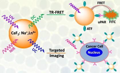
Ultrasmall Ln3+-doped CaF2 nanoprobes for TR-FRET detection of tumor marker suPAR and targeted cancer cell imaging (Image by Prof. CHEN Xueyuan's group).
Previously, Prof. CHEN's group had made a series of relevant progress on the controlled synthesis, optical physics, and bioapplications of Ln3+-doped nanoprobes. They reported the homogeneous TR-FRET detection of avidin with a detection limit of 4.8 and 5.5 nM, respectively, by employing Ln3+-doped NaYF4 and KGdF4 nanoprobes (Angew. Chem. Int. Ed. 2011, 50, 6306; J. Am. Chem. Soc. 2012, 134, 1323). Besides, they also developed a new inorganic oxide bioprobe based on sub-5 nm tetragonal ZrO2:Ln3+ NPs that could be used for sensitive detection of 3 nM of avidin and targeted cancer cell imaging (J. Am. Chem. Soc. 2012, 134, 15083).
Contact:
Prof. CHEN Xueyuan
Fujian Institute of Research on the Structure of Matter
Chinese Academy of Sciences
Email: xchen@fjirsm.ac.cn